テレメディカは、医療従事者と患者様の
よりよい信頼関係づくりのために
聴診教育に取り組んでいます
よりよい信頼関係づくりのために
聴診教育に取り組んでいます

【PR】PAS5.消化器系のアセスメント|講師 山内豊明|看護実践能力開発講座!フィジカルアセスメント

看護実践能力開発講座!フィジカルアセスメント
テーマ「5.消化器系のアセスメント」
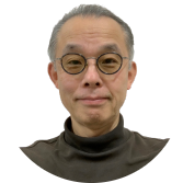
講師 山内豊明
放送大学大学院教授
名古屋大学名誉教授
ダイジェスト動画 2分31秒
■セミナーお申込み
https://telemedica.site/seminar-top/
■看護実践能力に基づく学習項目
この講座のテーマは、看護実践能力に基づく学習項目の下記に該当します。
能力:臨床実践能力
1.能力の構成要素:ニーズをとらえる力
学習項目:アセスメント(身体面)
2.能力の構成要素:ケアする力
学習項目:看護技術
3.能力の構成要素:意思決定を支える力
学習項目:看護・医療の方針等を話し合うプロセス
■セミナーを受講してデジタルバッチ(オープンバッチ)を獲得しよう!
本プログラムを両方修了された方に、習得したスキルを証明するためのデジタル修了証「オープンバッジ」を発行いたします。
© 2026 Telemedica, Inc. All rights reserved.
【PR】看護実践能力開発講座!フィジカルアセスメント|講師 山内豊明
実践!フィジカルアセスメントセミナーのご案内
Web+ハンズオンで、系統的に学び、実践で仕上げる。
講師 山内豊明
放送大学大学院教授
名古屋大学名誉教授
看護現場で求められるフィジカルアセスメント能力を、系統的かつ実践的に学べる講座です。
本講座は、全7回のWeb動画セミナー+ハンズオンセミナー1日で構成されています。
ダイジェスト動画(1分29秒)
動画で知識を身につけ、ハンズオンで実践することで、“わかる”を“できる”へつなげます。
各回の講義では、手元資料(講義資料)のダウンロードが可能。さらに、ハンズオン受講者はiPaxの聴診症例・聴くゾウを利用しながら、実践的な聴診トレーニングを行えます。
ハンズオンセミナー修了者には、デジタル修了証(オープンバッジ)を発行。受講した学びを、スキルの証明として記録・活用できます。
この講座の特長
✅ Webで自分のペースで学べる
✅ 講義資料をダウンロードして復習できる
✅ iPax聴診症例・クイズが利用できる
✅ ハンズオンで実技を習得できる
✅ 修了者にオープンバッジ発行
全7回 Web動画セミナーテーマ
第1回|看護実践に不可欠なフィジカルアセスメント
第2回|臨床推論の進め方・方法論 ~症状・徴候からのアプローチ~
第3回|呼吸器系のアセスメント
第4回|循環器系のアセスメント
第5回|消化器系のアセスメント
第6回|意識と中枢神経系のアセスメント
第7回|感覚器系・運動器系のアセスメント
詳細はこちら
https://telemedica.site/seminar-top/
© 2026 Telemedica, Inc. All rights reserved.
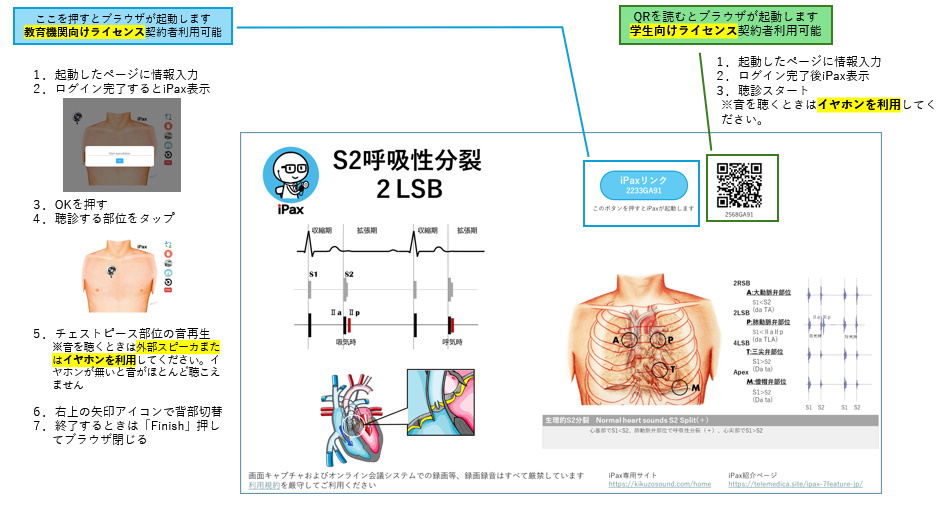
【PR】PPTで使う
音源の「症例リスト」一例
\ 合計700症例以上 /
心音(iPax症例)

・正常心音
・S2呼吸性分裂
・S2異常分裂
・S3ギャロップ
・S4ギャロップ
・サメーションギャロップ
・大動脈弁狭窄
・大動脈弁閉鎖不全
・僧帽弁狭窄
・僧帽弁閉鎖不全
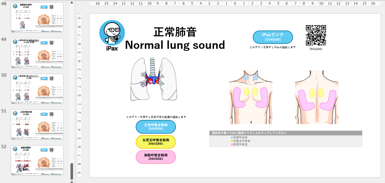
肺音(iPax症例)

・正常肺音
・ファインクラックルズ
・コースクラックルズ
・ウィージズ
・ロンカイ
・ウィージズ+ロンカイ
・コースクラックルズ+ロンカイ
・コースクラックルズ+スクウォーク +ロンカイ
・空洞呼吸
・気管支呼吸音低下
その他聴診音

・コロトコフ音
(脈触診機能あり含む)
・グル音
(メタリックサウンド含む)
・透析シャント音
ケーススタディ

・心不全
(僧帽弁閉鎖不全)
・心不全
(僧帽弁閉鎖不全+三尖弁閉鎖不全 +心房細動)
・胸痛の症例
・心電図異常がある症例
・MTX肺の関節リウマチ症例
・関節リウマチに伴う間質性肺炎 COPD合併例
・蜂巣肺のない特発性肺疾患症例
・高MDA-5抗体陽性皮膚筋炎症例
・新生児症例
\ リストに無い症例やオリジナル症例を希望の場合は、テレメディカが音源を作成して提供します /